TESTIMONIALS* –
CLINICAL CASES
The high-performance CBCT system from PreXion can be used in all areas of modern dentistry and beyond.
Case one

Sam J. Halabo, DMD
Chula Vista, CA, USA
* The testimonials refer to the PreXion Excelsior CBCT system, which is similar to the PreXion3D EXPLORER and already established in the US market.
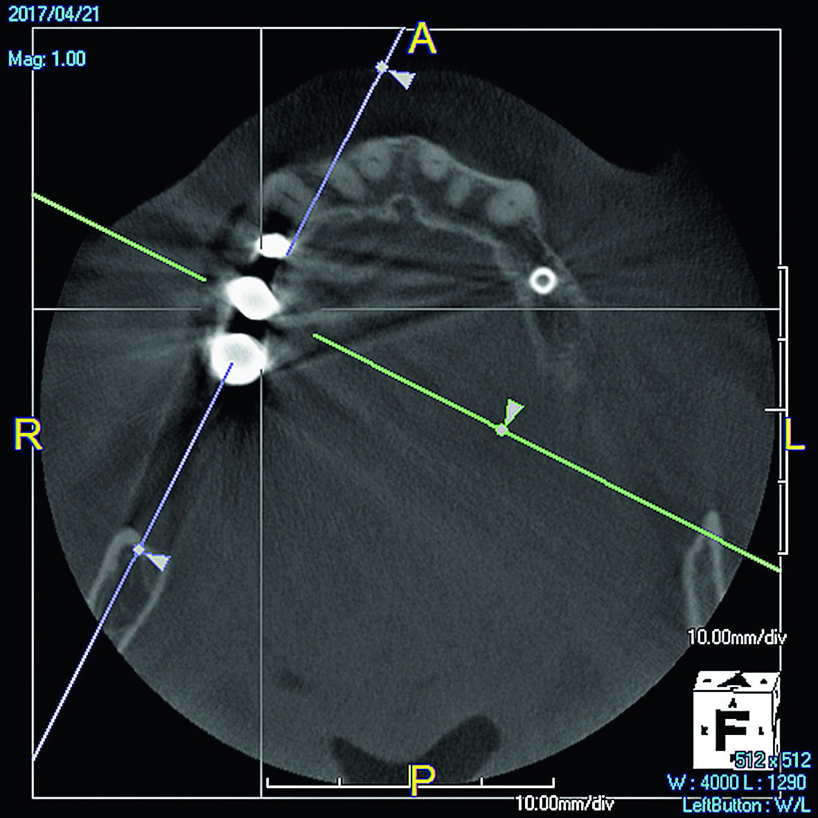
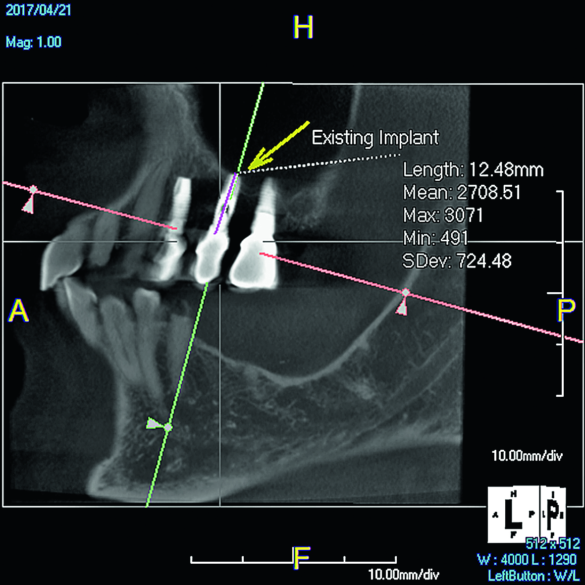
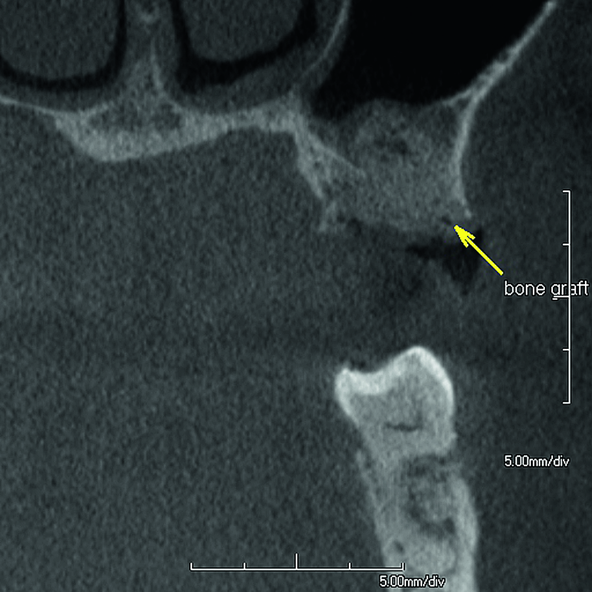
This scan is of a 68-year-old female who came to my practice for a full exam. When we examined her, we noted that there were implants on both the upper right and the upper left, underneath her fixed bridges. After taking a cone beam scan, we could clearly see that the existing implants were not part of her bridges, this was an unpleasant surprise to her. More importantly, we could see that the bridge on the upper right had failed and the one on the left was starting to as well. The patient agreed that it was important to immediately address the upper right side. The cone beam scan was essential in deciding how to treat the patient.
The biggest challenge in this case was trying to use the existing implant that had not been utilised since its initial placement over ten years ago. We first had to figure out the type and the size of that implant, as well as how to access it. The improved imaging and the measurement tool in the PreXion3D Viewer allowed me to do so easily and with accuracy. We removed the bridge and remaining failing teeth, performed a sinus lift, a diagnostic wax-up was made, we placed two implants in the adjacent areas, and finally restored to get her upper right side to a completely manageable state.
Case two

Jon M. Julian, DDS
Travelers Rest, SC, USA
* The testimonials refer to the PreXion Excelsior CBCT system, which is similar to the PreXion3D EXPLORER and already established in the US market.
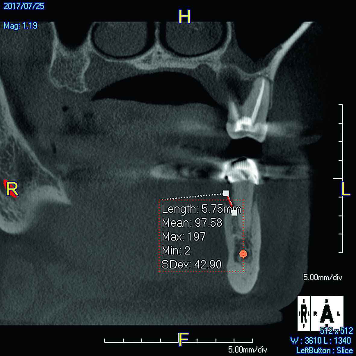
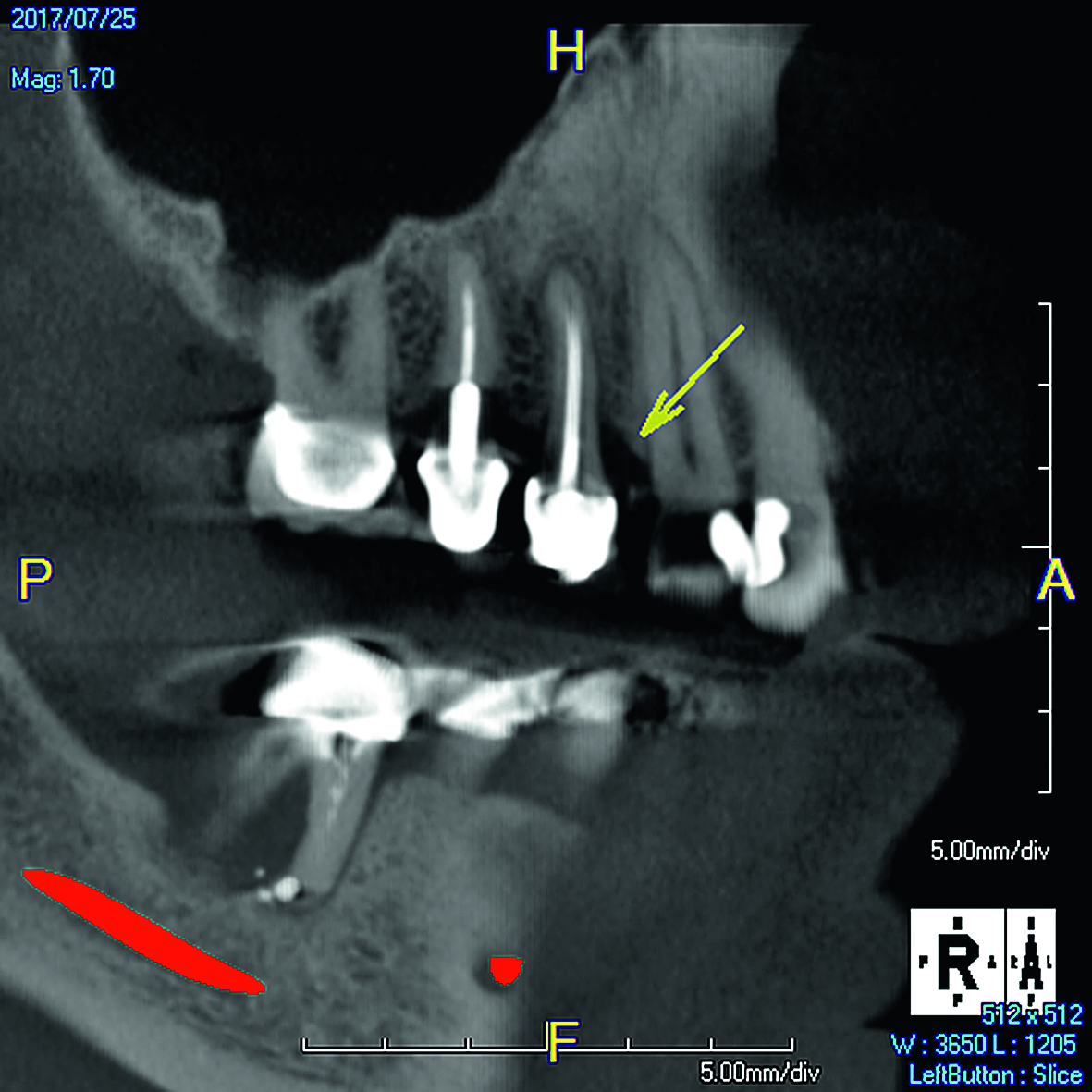
This 65-year-old patient came to our practice with upper left sore tooth pain. He initially went to his dentist three months previously for a routine oral care and hygiene visit. Now he was hesitant to take on more X-ray procedures, however, I asked him if he would be willing to take a beneficial cone beam scan at no charge. He agreed and proceeding forward we discovered on the cone beam imaging the source of his pain, as tooth #14 had a large radiolucency around all three roots. It was completely mobile and the patient fully understood the reason for its recommended removal in the course of treatment.
In addition we asked the patient if its okay to discuss the other findings displayed on the cone beam scan that he was unaware of or even existed. He agreed and what we found on the lower right was another abscess involving tooth #30 and his previous dentist evidently did not discover it with his 2D X-ray imaging. Finally we asked the patient if he knew anything about periodontal disease? He mentioned being unaware of it and didn’t think he possibly had any. To help explain we showed him a 9 mm pocket on the distal premolar, a 6 mm pocket on
the lingual pre-molar and a 5–6 mm pocket on an upper premolar. We were able to visually define and engage him in his own co-diagnosis along with a little education of the nature of periodontal/pathological processes in his own mouth. The patient agreed to all periodontal therapy and treatments beyond just the routine hygiene cleanings. I feel like I would never have been able to explain his situation properly without the visual cone beam images to demonstrate to the patient exactly the full scope of his situation even though his only pain was present in the upper left tooth #14.