TOP


Flat panel detector
By irradiating X-ray on the internal scintillator, the emitted light is accumulated inside as electrical signal and will be output as digital data.
Handgrip
A handgrip for the patient to grab.
Status indicator
It shows scanner status.
X-ray tube head
Generates the X-ray beam required for scanning.
Chin rest
A place to put the patient‘s chin on.
Vertical laser window
Laser beam is irradiated while positioning a patient.


Why 2D + 3D?
Three-dimensional CBCT imaging is the decisive advantage compared to conventional 2D X-ray equipment, as the dentist can spatially examine the oral conditions according to the most varied medical aspects. Compared to the three-dimensional image information obtained with a CBCT scan and that of a 2D X-ray image, the radiation exposure of the latter is disproportionately high. In addition, the volume structure of the hard and soft tissue is incomparably better represented than in 2D X-rays.
Why a large FOV?
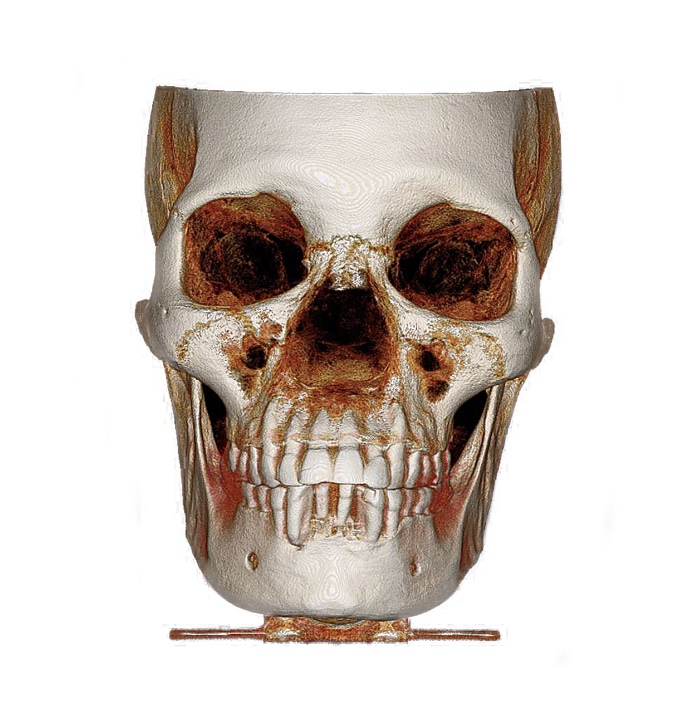
With one of the largest field of views (FOV) on the market (150 x 160 mm), the PreXion3D EXPLORER can display all the important anatomical structures of the skull in detail. The powerful imaging software helps to highlight and measure relevant areas. Particularly in oral and maxillofacial surgery as well as ear, nose and throat medicine, large-area spatial image analysis helps to develop the best therapy options.
Why PreXion3D EXPLORER?
The powerful system components of the PreXion3D EXPLORER enable an extraordinary combination of the most precise 3D imaging, large image detail, lowest radiation exposure, reliable diagnostics and digital planning for all indications in modern dentistry such as periodontology, endodontics, implan-tology or maxillofacial surgery. The so-called patient management system is designed for secure and networked communication of patient data across the various practice rooms and can be integrated into the existing infrastructure. With the precision and professional competence of PreXion, dentists have the right partner at their side.


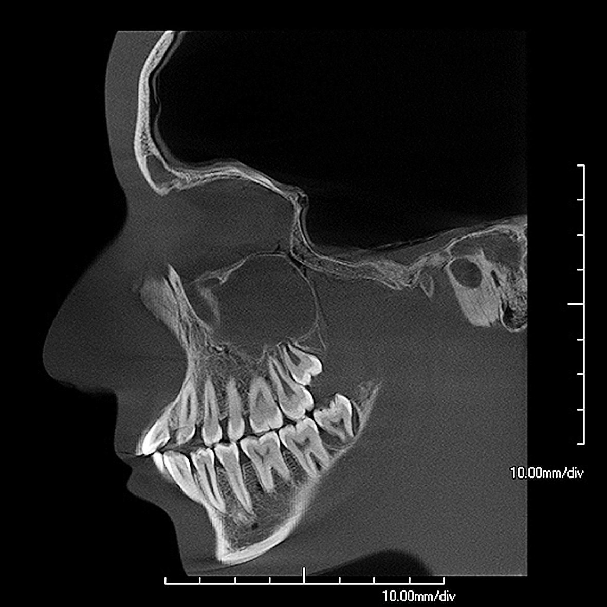
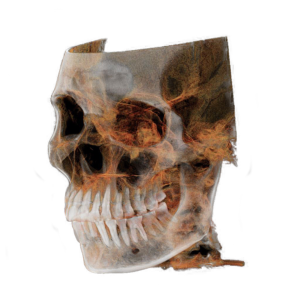
soft tissue
Successful therapy requires, among other things, a detailed representation of the soft tissue, the head and facial nerves.
hard tissue
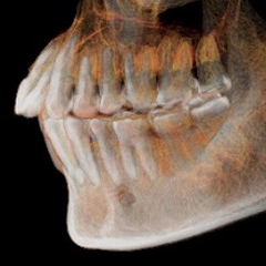
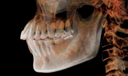
The voxel size of only 0.075 mm allows the different bone densities of the skull to be precisely represented.
With many 3D-imaging systems today, good image quality is usually accompanied by high radiation exposure. The PreXion3D EXPLORER, offers with a 0.3 mm focal spot and voxel size of 0.075 mm a unique combination of highest possible image quality with lowest possible radiation exposure.


soft tissue
Successful therapy requires, among other things, a detailed representation of the soft tissue, the head and facial nerves.
hard tissue
The voxel size of only 0.075 mm allows the different bone densities of the skull to be precisely represented.
The output in ultra-HD with a small voxel size enables a more detailed representation of even the finest structures.
Voxelsize 100 µm

Voxelgröße 75 µm

Fokuspunkt 0.5 mm

Fokuspunkt 0.3 mm
Focal spot of the X-ray beam
The focal spot is the area on the target of the X-ray tube which the electron stream strikes and from which X-rays are emitted. It is also called focus. The larger the area of the focal spot, the poorer is the detail in the X-ray image. The PreXion3D EXPLORER has a 0.3 mm focal spot. One of the smallest in the industry.

What is voxel?
It is a 3D volume element shaped as an isotropic cube. Hounsfield units are used as a quantitative measurement of radio density in voxel. Voxel is a combination of the words volumetric and pixel.

The PreXion3D EXPLORER provides an accurate 360° panoramic view from 512 to 1,024 projected views. In addition to the 3D analysis function for image detail sizes (FOV) of 50 x 50, 150 x 80 and 150 x 160 mm, it has a “True” and a “Reconstructed” panorama mode. The device impresses with its ease of operation, comprehensive planning programmes and imaging software across all dental indication areas.

50×50
100×100
150×80

150×160

Scanner connection
The color of the display changes depending on the scanner status:
Patient data
Preferences/Positioning

3D-navigation
Analysis
Endodontic tools
Implant tools
Interfaces